Anatomy Muscles Pelvis : Hip Pain Explained Including Structures Anatomy Of The Hip And Pelvis - It helps maintain erect posture, abducts the thigh, and rotates the thigh outward.
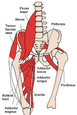
Anatomy Muscles Pelvis : Hip Pain Explained Including Structures Anatomy Of The Hip And Pelvis - It helps maintain erect posture, abducts the thigh, and rotates the thigh outward.. The floor of the pelvis is made up of the muscles of the pelvis, which support its. The three muscles of the group—the iliacus, the psoas major, and the psoas minor—arise from different areas of your pelvis and lumbar spine to form a common attachment in your hip. Pelvic floor muscles located wholly within the pelvis The muscles of the abdomen, lower back, and pelvis are separated from those of the chest by the muscular wall of the diaphragm, the critical breathing muscle. Any injury or disease of the hip will adversely affect the joint's range of motion and ability to bear weight.
Use the mouse scroll wheel to move the images up and down alternatively use the tiny arrows (>>) on both side of the image to move the images.>>) on both side of the image to move the images. The four groups are the anterior group, the posterior group, adductor group, and finally the abductor group. The pelvis consists of the sacrum, the coccyx, the ischium, the ilium, and the pubis. Über 7 millionen englischsprachige bücher. It originates from the pelvic outermost layer of the middle 3 sections of sacrum by 3 digitations.
Pelvis Wikipedia from upload.wikimedia.org The main focus of this article will be the pelvic floor muscles.on that topic, there are several important questions that need to be answered: The structure of the pelvis supports the contents of the abdomen while also helping to transfer the weight from the spine to the lower limbs. The classification of the two groups under a common heading is. Each hip bone, in turn, is firmly joined to the axial skeleton via its attachment to the sacrum of the vertebral column. The ligaments, made of strong connective tissue, which connect bones to bones, and the tendons, which connect muscles to bones. The pelvis contains a large number of organs, bones, muscles, and ligaments, so many conditions can affect the entire pelvis or parts within it. The largest of them is the most superficial muscle, the gluteus maximus. The muscles of the abdomen, lower back, and pelvis are separated from those of the chest by the muscular wall of the diaphragm, the critical breathing muscle.
The ilium, ischium and the pubic bone.
Large ligaments, tendons, and muscles around the hip joint hold the bones (ball and socket) in place and keep it from dislocating. The iliopsoas muscle is a major hip flexor that also helps to move your spine. The floor of the pelvis is made up of the muscles of the pelvis, which support its. Any injury or disease of the hip will adversely affect the joint's range of motion and ability to bear weight. The ilium, ischium and the pubic bone. (2) the levator ani and the coccygeus, which together form the pelvic diaphragm and are associated with the pelvic viscera. The structure of the pelvis supports the contents of the abdomen while also helping to transfer the weight from the spine to the lower limbs. The pelvic floor muscles include; The muscles within the pelvis may be divided into two groups: The anterior muscle group features muscles. It originates from the pelvic outermost layer of the middle 3 sections of sacrum by 3 digitations. Similar to learning the muscles of the lumbar spine/trunk, it can be helpful to first look at the. Cross the ls joint onto the trunk 2.
The pelvic floor is primarily made up of thick skeletal muscles along with nearby ligaments and their investing fascia. The pelvis consists of the sacrum, the coccyx, the ischium, the ilium, and the pubis. Name given to a group of muscles. It originates from the pelvic outermost layer of the middle 3 sections of sacrum by 3 digitations. (1) the obturator internus and the piriformis, which are muscles of the lower extremity, and will be described with these (pages 476 and 477);
Pelvis Hip Anatomy from uploads-ssl.webflow.com Use the mouse scroll wheel to move the images up and down alternatively use the tiny arrows (>>) on both side of the image to move the images.>>) on both side of the image to move the images. Some conditions that can affect the female pelvis. The pelvic floor is primarily made up of thick skeletal muscles along with nearby ligaments and their investing fascia. Cross the ls joint onto the trunk 2. The bones of the pelvis are held together by a large number of ligaments and muscles. It is usually divided into two separate anatomic regions: Structures arteries of the pelvis and perineum bones of the pelvis and perineum fascia of the pelvis and perineum joints of the pelvis and perinium lymphatics of the pelvis and perineum muscles of the pelvis and perineum nerves of the pelvis and perineum topographical anatomy of pelvis and perineum veins… (2017, elsevier) should be consulted.
The pelvic girdle and pelvic spine.
The pelvic diaphragm is the third deepest layer of the pelvic floor which puts it at the very center of all the other muscles. Lying exposed between the protective bones of the superiorly located ribs and the inferiorly located pelvic girdle, the muscles of this region play a critical role in protecting the. Knowing the anatomy of this muscle can help you make good choices in caring for an. It's supplied by ventral rami of first and 2nd sacral nerves (s1, s2). It is also referred to as a ball and socket joint and is surrounded by muscles, ligaments, and tendons. It separates pelvis from the perineum. The classification of the two groups under a common heading is. The pelvis consists of the sacrum, the coccyx, the ischium, the ilium, and the pubis. The pelvic girdle (hip girdle) is formed by a single bone, the hip bone or coxal bone (coxal = hip), which serves as the attachment point for each lower limb. Supports and positions the pelvic organs. Name given to a group of muscles. The pelvic floor is primarily made up of thick skeletal muscles along with nearby ligaments and their investing fascia. The pelvic floor muscles include;
Pelvic floor muscles located wholly within the pelvis The pelvic girdle and pelvic spine. Posterior pelvic ring fractures of the si joint and the sacrum ortho illinois pelvic anatomy the hip is a ball and socket joint in which the head of the femur the leg bone fits into the pelvis. (2) the levator ani and the coccygeus, which together form the pelvic diaphragm and are associated with the pelvic viscera. It's supplied by ventral rami of first and 2nd sacral nerves (s1, s2).
Pelvis Wikipedia from upload.wikimedia.org Cross the hip joint onto the thigh/leg 3. It is also referred to as a ball and socket joint and is surrounded by muscles, ligaments, and tendons. The muscles within the pelvis may be divided into two groups: Knowing the anatomy of this muscle can help you make good choices in caring for an. The pelvic floor is primarily made up of thick skeletal muscles along with nearby ligaments and their investing fascia. It is subdivided into the pelvic girdle and the pelvic spine. It is a broad flat muscle. The pelvic diaphragm is the third deepest layer of the pelvic floor which puts it at the very center of all the other muscles.
The pelvic girdle (hip girdle) is formed by a single bone, the hip bone or coxal bone (coxal = hip), which serves as the attachment point for each lower limb.
The structure of the pelvis supports the contents of the abdomen while also helping to transfer the weight from the spine to the lower limbs. How can you strengthen them? The main focus of this article will be the pelvic floor muscles.on that topic, there are several important questions that need to be answered: It is a broad flat muscle. The classification of the two groups under a common heading is. The pelvic girdle, also known as the hip bone, is composed of three fused bones: The right and left hip bones also converge anteriorly to attach to each other. The pelvic girdle (hip girdle) is formed by a single bone, the hip bone or coxal bone (coxal = hip), which serves as the attachment point for each lower limb. The piriformis is a triangular muscle 1 on either side on the very front of the posterior wall of true pelvis. The four groups are the anterior group, the posterior group, adductor group, and finally the abductor group. The muscles of the abdomen, lower back, and pelvis are separated from those of the chest by the muscular wall of the diaphragm, the critical breathing muscle. An important group of muscles in the pelvis is the pelvic floor.the pelvic floor muscles provide foundational support for the intestines and bladder. Knowing the anatomy of this muscle can help you make good choices in caring for an.


0 Komentar